Pass4Future also provide interactive practice exam software for preparing ARDMS Abdomen Sonography Examination (AB-Abdomen) Exam effectively. You are welcome to explore sample free ARDMS AB-Abdomen Exam questions below and also try ARDMS AB-Abdomen Exam practice test software.
Do you know that you can access more real ARDMS AB-Abdomen exam questions via Premium Access? ()
What is the normal Doppler waveform signature of the hepatic veins?
Answer : C
The normal hepatic vein Doppler waveform is triphasic, reflecting cardiac cycle variations in central venous pressure transmitted from the right atrium through the IVC. Loss of triphasicity may suggest elevated right atrial pressures or hepatic venous obstruction.
According to Rumack's Diagnostic Ultrasound:
''The normal hepatic vein waveform is triphasic due to transmitted right atrial pressure variations.''
Rumack CM, Wilson SR, Charboneau JW, Levine D. Diagnostic Ultrasound. 5th ed. Elsevier, 2017.
AIUM Practice Parameter for Abdominal Vascular Ultrasound, 2020.
Which condition is demonstrated in this image of the groin?

Answer : D
The ultrasound image demonstrates bowel loops with peristalsis visualized within the inguinal canal, which is diagnostic of an inguinal hernia---more specifically, an indirect inguinal hernia. Indirect hernias pass through the deep inguinal ring and may extend into the scrotum, appearing sonographically as bowel-containing masses adjacent to or within the scrotal sac. Peristaltic motion confirms the presence of viable bowel content.
This finding is typical in indirect inguinal hernias, which are more common in males and often congenital due to a patent processus vaginalis. The herniated bowel can be traced through the inguinal canal, as seen in this image.
Comparison of answer choices:
A . Hematocele presents as a complex fluid collection surrounding the testis, often due to trauma---no complex fluid or trauma is apparent here.
B . Testicular rupture shows discontinuity of the tunica albuginea and irregular testicular contour---none of which is seen.
C . Orchiectomy would show an absent testis---this is not the case here.
D . Indirect hernia is correct. The presence of bowel with peristalsis in the inguinal canal is diagnostic.
Rumack CM, Wilson SR, Charboneau JW, Levine D. Diagnostic Ultrasound, 5th ed. Elsevier; 2017.
AIUM Practice Parameter for the Performance of Scrotal Ultrasound Examinations (2021).
Dogra VS, Gottlieb RH, Rubens DJ, Oka M. Sonography of the scrotum. Radiology. 2003;227(1):18--36
Which patient maneuver would best aid in identifying the pathology demonstrated in this image?

Answer : D
The ultrasound image demonstrates a classic example of ascites, shown by the anechoic (dark) fluid located between bowel loops or surrounding abdominal organs. In this case, there appears to be a small fluid collection in the peritoneal cavity.
One of the key maneuvers used to differentiate free fluid (such as ascites) from loculated fluid or other structures is to reposition the patient. Asking the patient to ''turn from side to side'' (Option D) can help in assessing whether the fluid shifts position --- a hallmark feature of free intraperitoneal fluid. This positional change is highly useful in confirming the diagnosis and distinguishing ascites from other potential mimics (e.g., cystic masses, lymphoceles, or bowel wall thickening).
In contrast:
Drinking water (A) is often used in imaging the urinary bladder or gastrointestinal tract but not for fluid characterization.
Standing upright (B) may shift fluid but is less practical during real-time ultrasound.
Breathing quietly (C) doesn't significantly aid in visualizing peritoneal fluid mobility.
Rumack CM, Wilson SR, Charboneau JW, Levine D. Diagnostic Ultrasound, 5th ed. Elsevier; 2017.
Hagen-Ansert SL. Textbook of Diagnostic Sonography, 8th ed. Elsevier; 2017.
AIUM Practice Parameter for the Performance of Diagnostic and Screening Ultrasound Examinations of the Abdomen and/or Retroperitoneum (2020).
Which sonographic finding is commonly associated with transitional cell cancer of urinary bladder?
Answer : A
Transitional cell carcinoma (TCC) typically presents as a non-mobile, polypoidal, focal intraluminal mass projecting from the bladder wall. Mobility of the lesion helps differentiate TCC from blood clots or debris.
According to Rumack's Diagnostic Ultrasound:
''Bladder TCC most often appears as a non-mobile, polypoid mass attached to the bladder wall.''
Rumack CM, Wilson SR, Charboneau JW, Levine D. Diagnostic Ultrasound. 5th ed. Elsevier, 2017.
AIUM Practice Parameter for Bladder Ultrasound, 2020.
---
Which congenital anomaly is demonstrated in this image?

Answer : D
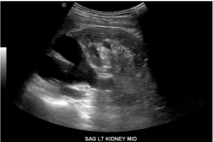
The ultrasound image labeled ''SAG LT KIDNEY MID'' demonstrates a left kidney with two separate, centrally located echogenic renal sinuses separated by intervening renal parenchyma. This appearance is classic for a duplicated collecting system.
A duplicated collecting system (also known as duplex kidney) is a congenital anomaly in which a single kidney contains two separate pelvicalyceal systems. It may be complete (with two ureters) or incomplete (partial duplication of ureters). This condition is one of the most common congenital anomalies of the urinary tract.
Sonographic Features of a Duplicated Collecting System:
Two separate central echogenic renal sinus regions seen within one kidney
Intervening parenchyma between the two sinuses
May show associated findings: hydroureteronephrosis (especially of upper pole moiety), ureterocele
Best visualized in sagittal plane
Differentiation from other options:
A . Horseshoe kidney: Shows fusion of the lower poles of the kidneys, typically located anterior to the aorta in the midline---not demonstrated here.
B . Crossed renal ectopia: One kidney is located on the opposite side of the body; this image shows a normally positioned kidney.
C . Prominent renal column (column of Bertin): May mimic a mass, but does not produce two separate sinuses as shown here.
Rumack CM, Wilson SR, Charboneau JW, Levine D. Diagnostic Ultrasound. 5th Edition. Elsevier, 2018. Chapter: Kidneys, pp. 210--215.
American Institute of Ultrasound in Medicine (AIUM). Practice Parameter for the Performance of a Renal Ultrasound Examination, 2020.
Radiopaedia.org. Duplex collecting system: https://radiopaedia.org/articles/duplex-collecting-system