Free Practice Questions for ARDMS AE-Adult-Echocardiography Exam
Pass4Future also provide interactive practice exam software for preparing ARDMS AE Adult Echocardiography Examination (AE-Adult-Echocardiography) Exam effectively. You are welcome to explore sample free ARDMS AE-Adult-Echocardiography Exam questions below and also try ARDMS AE-Adult-Echocardiography Exam practice test software.
Page: 1
/ 14 Total 139 questions
Do you know that you can access more real ARDMS AE-Adult-Echocardiography exam questions via Premium Access? ()
Question 1
How must the sonographer angle the transducer from the apical four-chamber view in order to visualize the aortic valve in the apical five-chamber view?
Answer : A
To obtain the apical five-chamber view from the apical four-chamber, the transducer is angled anteriorly (towards the patient's chest). This brings the left ventricular outflow tract and aortic valve into the imaging plane anterior to the left ventricle and mitral valve seen in the four-chamber view.
Posterior, medial, or lateral angulations do not adequately visualize the aortic valve in this context.
This technique is described in adult echocardiography imaging protocols and ASE chamber quantification guidelines12:ASE Imaging Protocolsp.30-3516:Textbook of Clinical Echocardiography, 6ep.70-75.
Question 2
Which finding occurs initially as the seventy of aortic stenosis progresses?
Answer : A
In the early stages of aortic stenosis, the left ventricle adapts to increased afterload by concentric remodeling, which is characterized by increased wall thickness without a proportional increase in chamber size. This adaptation helps normalize wall stress.
As the disease progresses, concentric hypertrophy develops with thickened walls and decreased compliance. Eccentric hypertrophy and global systolic dysfunction occur later with decompensation and ventricular dilation.
This progression is explained in the 'Textbook of Clinical Echocardiography, 6e', Chapter on Left Ventricular Adaptations to Pressure Overload20:365-370Textbook of Clinical Echocardiography.
Question 3
How is the aorta in a structurally normal heart oriented?
Answer : B
Compreh ensive and Detailed Explanation From Exact Extract:
In a normal heart, the ascending aorta arises from the left ventricle and courses superiorly and posteriorly to the right of the pulmonary artery, which arises anteriorly from the right ventricle. The aorta is positioned posterior and to the right of the main pulmonary artery, reflecting the normal spatial relationship.
The pulmonary artery is anterior and to the left of the aorta, and the coronary sinus lies posteriorly in the atrioventricular groove.
This anatomical relationship is detailed in the 'Textbook of Clinical Echocardiography, 6e', Chapter on Cardiac Anatomy and Echocardiographic Landmarks20:50-55Textbook of Clinical Echocardiography.
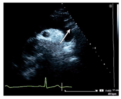
Question 4
Which artery is identified by the arrow on this image?
Answer : B
The image is a suprasternal or high parasternal echocardiographic view of the aortic arch and its branches. The arrow points to the first large branch arising from the aortic arch, which is the brachiocephalic artery (also called the innominate artery). This vessel courses superiorly and bifurcates into the right common carotid and right subclavian arteries.
The left common carotid artery is the second branch from the arch, the left subclavian artery is the third branch, and the right common carotid is a branch of the brachiocephalic artery, not directly off the arch.
This anatomic arrangement and its echocardiographic depiction are well documented in adult echocardiography references and vascular ultrasound guidelines12:ASE Vascular Imaging Guidelinesp.270-27516:Textbook of Clinical Echocardiography, 6ep.400-405.
Question 5
The 'P' wave of an electrocardiogram relates to which echocardiography event?
Answer : A
Comprehensive and Detailed Explanation From Exact Extract:
The P wave on the ECG corresponds to atrial depolarization, which precedes atrial contraction (atrial systole). On echocardiography, atrial contraction can be observed as the atrial 'kick,' contributing to ventricular filling during late diastole.
Ventricular contraction (QRS complex) and ventricular relaxation (T wave) correspond to other phases of the cardiac cycle. Atrial relaxation occurs during ventricular systole but is not represented by the P wave.
This timing relationship is critical for correlating echocardiographic Doppler inflow patterns, such as the late diastolic A wave, with the ECG. These concepts are outlined in the foundational echocardiography references, including ASE guidelines and the 'Textbook of Clinical Echocardiography'16:Textbook of Clinical Echocardiography, 6ep.150-15512:ASE Echocardiography Guidelinesp.50-55.
Page: 1
/ 14 Total 139 questions
Unlock Full AE-Adult-Echocardiography Exam Features